What Are Stretch Marks?
Definition, epidemiology, characteristics, reasons, localisations and treatments for stretch marks
Stretch marks, commonly said “striae distensae”, are real skin lesions which appear in different shape and colour in relation to their evolution.
The cause of the striae formation has not yet been entirely clarified, it is considered to be the consequences of mechanical stress, such as weight and volume gain or hormonal changes which characterize e.g. puberty, pregnancy and breastfeeding, or hormonal or cortisone treatments or particularly intense physical activity [01, 02].
Many times, some of the causes occur simultaneously, so much so that the most recent research speaks of multifactorial causes 02. Stretch marks are sometimes ominous symptoms of serious diseases such as Marfan Syndrome [03] or Cushing Syndrome [04]; the University of Verona has recently discovered that the stretch marks can be a symptom which allows to diagnose forms of autism in children before they even show themselves [05].
The most affected areas are the breasts, abdomen, gluteus and thighs in women, while in men they generally show up on the shoulders and deltoids, kidney and hip areas 06. From a structural point of view, we know that stretch mark is a consequence of a collagen alteration which frays and no longer offers support to our skin [03].
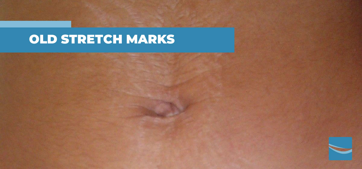
The first phase in the life of a stretch mark, when specified as “striae rubra” or “red stretch mark”, corresponds to an inflammatory state04 which can extend up to two years from its onset. During this period, they appear red and noticeable and are characterised by an intense colour and sometimes can be itchy. Stretch marks progressively change in appearance and colour.
The intense red colour becomes progressively lighter than the surrounding tissue and later tends to take an opaque tonality. Therefore, these stretch marks are defined as ‘striae albae’ or ‘white stretch marks’ and become evident during the so-called ‘scarring phase’.
At the same time, the stretch mark empties out progressively, causing tissue atrophy and showing up empty and deeper, becoming more evident both to the touch and to the eye.
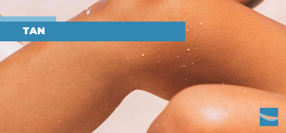
When the stretch mark turns white, it loses its ability to tan, because in time the basal membrane, in other words the thin layer which separates the epidermis from the dermis, becomes completely destructed01, losing melanocytes which means the elements that produce melanin with the sun exposure [07].
This makes stretch marks an aesthetic condition that is particularly prevalent in countries where sun exposure is recurrent during the warm season.
In fact, the tanning of the healthy skin surrounding the stretch marks, which remain white and opaque, makes them much more visible, resulting in what is called ‘zebra effect’ which makes them look particularly unpleasant to the eye.
In the first half of 20th century stretch mark was a quite rare phenomenon which sometimes affected women during pregnancy, specifically the first one. Around the end of the century, this alteration increased its effect reaching out to more than %60 of women after puberty, when it began to show itself significantly.
During the 21st century stretch mark progressively increased its effect to affect between %80 and 90% of women in the post-puberty age and began to be common even in young people starting from the time of development.
For a very long time it was considered that stretch mark was a cutaneous disease that was impossible to obtain significant improvements: the scientific literature in fact proved that it is possible to cure it successfully when the stretch mark is recent and red, while with the old, white and opaque stretch mark, the results were insufficient and occasional.
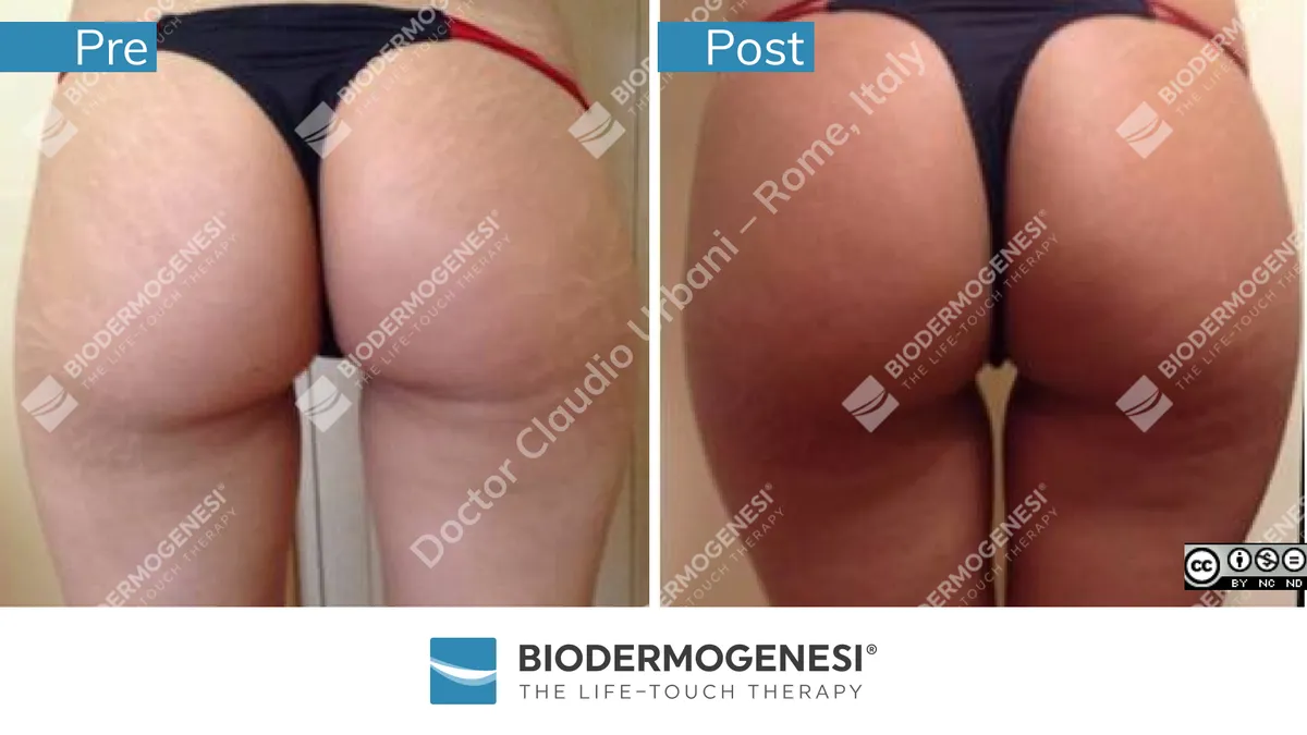
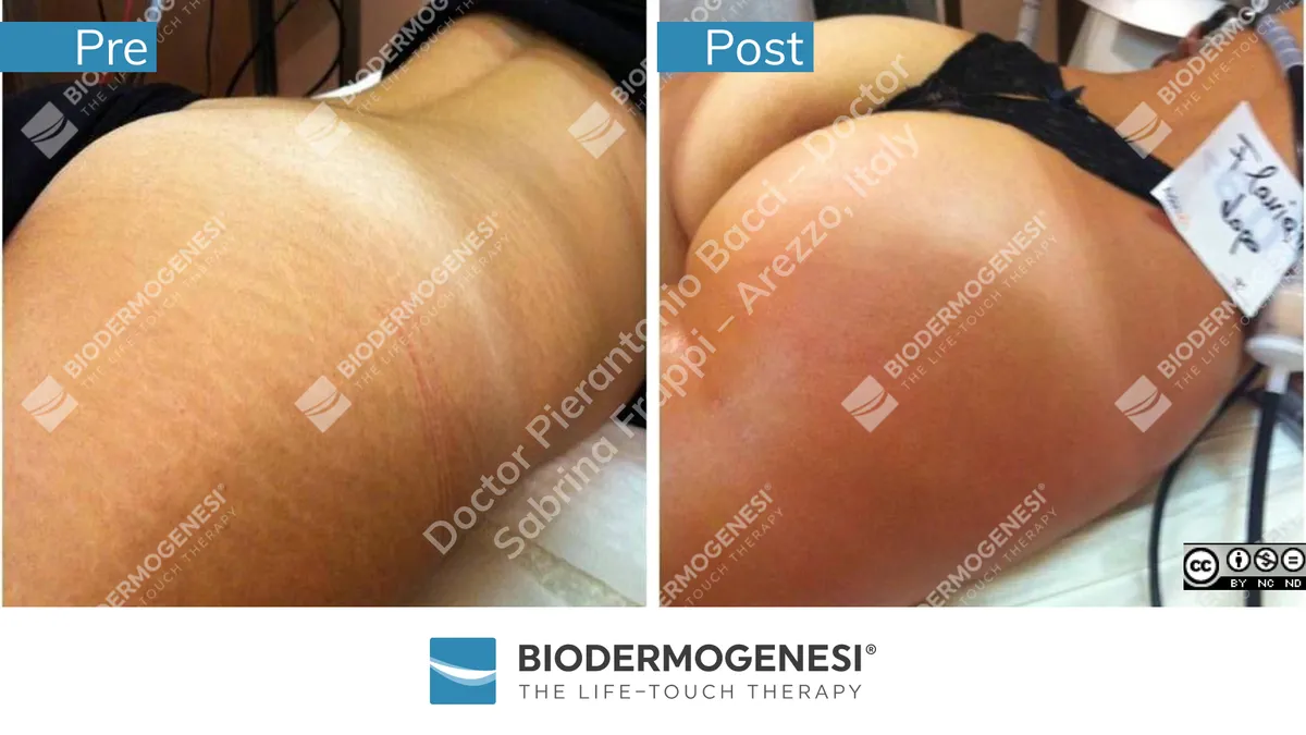
Recently Biodermogenesi®, a new medical method completely developed in Italy, has proved to regenerate both white and red stretch marks [07, 08, 09, 10, 11].
A study developed by three Italian universities07 demonstrated that Biodermogenesi® regenerates stretch marks, filling them but also reorganising them like the adjacent healthy skin, reorganising the basal membrane and reproducing melanocytes that will allow to have a normal tan of the stretch marks that can be obtained even with white stretch marks older than 25 years [09, 10].
Thanks to the total lack of side effects, Biodermogenesi® has proven to be extremely effective and also safe for the patient [07, 08, 09, 10, 11].
The pleasure of the therapy, the absence of downtime and the new-found ability to tan stretch marks make Biodermogenesi® patient-friendly.
References:
- Adam Hague, Ardeshir Bayat. Therapeutic targets in the management of striae distensae: A systematic review. J Am Acad Dermatol Vol 77, number 3, September 2017:559-568
http://dx.doi.org/10.1016/j.jaad.2017.02.048 - Raquel Cristina Tancsik Cordeiro, Aparecida Machado de Moraes. Striae Distensae: physiopathology. Surgical & Cosmetic Dermatology 2009;1(3):137-140.
https://www.researchgate.net/publication/286949690_Striae_distensae_Physiopathology - H Pinkus, M K Keech, H Mehregan. Histopathology of striae distensae, with special reference to striae and wound healing in the Marfan Syndrome. The Journal of Investigative Dermatology Vol. 46 n. 3 283-292 DOI: 10.1038/jid.1966.43
- H M Sheu, H S Yu, C H Chang. Mast cell degranulation and elastolysis in the early stage of striae distensae. J Cutan Pathol. 1991 Dec;18(6):410-6. doi: 10.1111/j.1600-0560.1991.tb01376.x
- Sheila Veronese, Leonardo Zoccante, Nicola Smania, Andrea Sbarbati. Stretch marks: a visible expression of connective’s involvement in autism spectrum disorders. Front. Psychiatry 14:1155854. doi: 10.3389/fpsyt.2023.1155854
- Francesca de Angelis, Larissa Kolesnikova, Franco Renato, Giuseppina Liguori. Fractional Nonablative 1540-nm Laser Treatment of Striae Distensae in Fitzpatrick Skin Types II to IV: Clinical and Histological Results. Aesthetic Surgery Journal 31(4) 411–419 DOI: 10.1177/1090820X11402493
- Antonio Scarano, Andrea Sbarbati, Roberto Amore, Eugenio L. Iorio, Giuseppe Ferraro, Felice Lorusso, Domenico Amuso. A New Treatment for Stretch Marks and Skin Ptosis with
Electromagnetic Fields and Negative Pressure: A Clinical and Histological Study. J Cutan Aesthet Surg. 2021 Apr-Jun; 14(2): 222–228. doi: 10.4103/JCAS.JCAS_122_20: 10.4103/JCAS.JCAS_122_20 - Artigiani A, Cervadoro G, LogginiB, Paolicchi A. Biodermogenesi®: la soluzione non invasiva nel trattamento delle smagliature. La Medicina Estetica 2012; 1,41-49
- Alberti G, Laura S. Treatment of stretch marks aged more than twenty years with the synergy of electromagnetic field and vacuum. Clinical case studies and subsequent follow up. Aesthetic Medicine 2019; 5(1)14-21.
- Bacci PA, Alberti G, Amuso D, Artigiani A, Benitez Roig V, Di Nardo V, Garcia-Gimenez V, Greco D, Laura S, Pagano M, Reale A, Sarracco I, Saracoglu S, Urbani C, Venditti E, Wade M, Zunica R. The synergy between vacuum and electromagnetic fields in the treatment of striae distensae: retrospective study on 917 patients with clinical and histological case records A possible treatment for striae distensae. Journal of Applied Cosmetology, Vol. 39 iss. 1 (January-June, 2021): 43-54. ISSN 0392-8543
- Veronese S, Bacci PA, Garcia Gimenez V, Canel Micheloud CC, Haro García NL, Sbarbati. A. V-EMF therapy: A new painless and completely noninvasive treatment for striae gravidarum. J Cosmet Dermatol. 2024;00:1-8. doi:10.1111/jocd.16220