Description and definition of scar
Scars differ from each other for different features, let’s learn to know them.
What are scars?
Scars are lesions of our skin that sometimes also affect our inner organs, such as c-section scars where skin layers, muscles and uterus are severed, or such as appendicectomy where skin layers and muscles are cut.
Many are the causes of these wounds: surgery, abrasion, cut, trauma, fire or heat, cold or chemical agents.
Scarring is a healing process activated by our organism to close the lesion as soon as possible and prevent death due to excessive blood loss or infection; for this reason, our body generates new imperfect tissue but not its skin appendages such as hair.
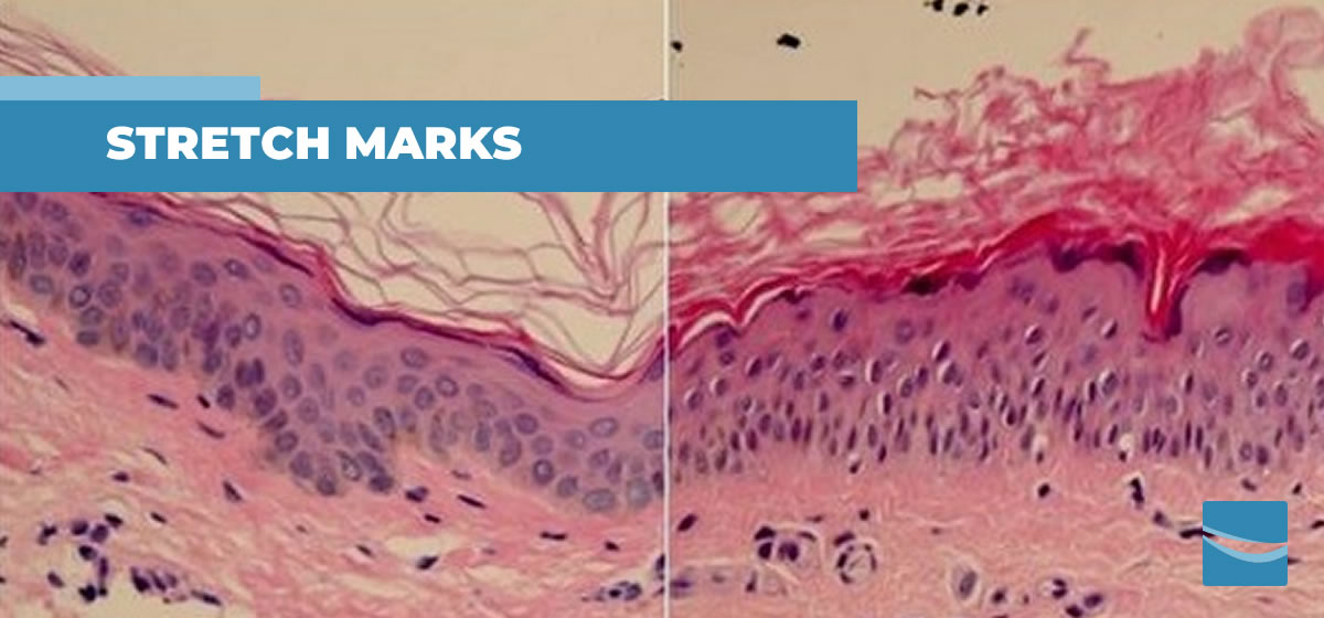
What do scars look like?
Scars’ texture is usually smooth and do not match the color of the surrounding healthy skin; it may be lighter or darker, tending to wine red color especially in presence of keloids.
Sunken and hollow scars are known as hypotrophic scars, whilst raised ones are called hypertrophic. Some scars grow larger than the injury that caused it and raise significantly, these take the name of keloids.
Unsightly appearance and functional consequences
Sometimes scars do not concern only the unaesthetic appearance but also have functional repercussions that may limit head, neck, shoulders and arms motility, or cause occasional/chronic pain.
These limitations are due to retraction of the scar tissue and formation of fibrosis inside. Fibrosis is compacted and hardened tissue that progressively extends, highly impacting the quality of the life of the patient.
Scientific research
Scientific research teaches us that the risk of scars degenerating into hypertrophic and keloids as well as of fibrosis forming are the consequences of an inflammatory state of the tissue (01, 02) or loss of hydration of the scar (03, 04).
In conclusion
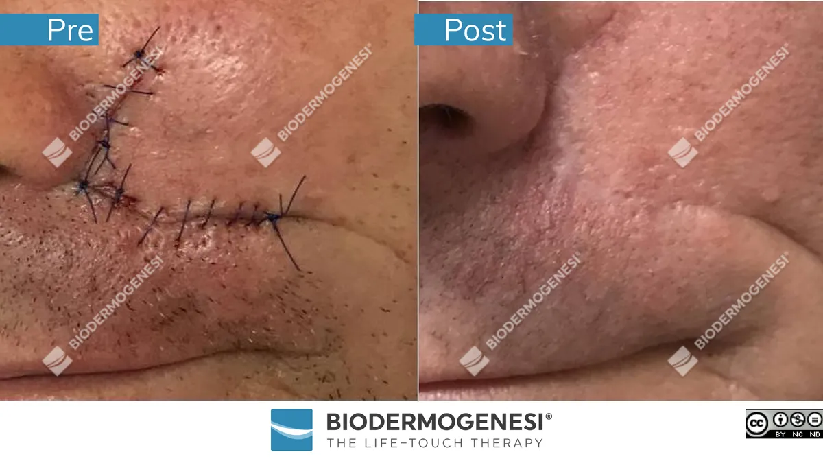
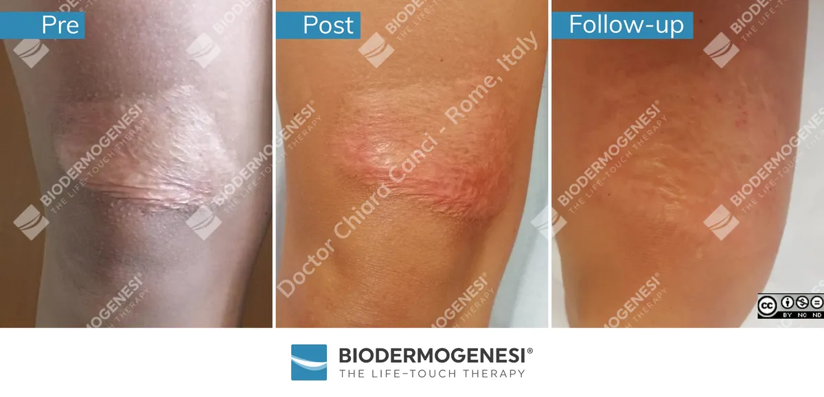
For these reasons it is useful to cover the scars with biocompatible silicon film (05) as it allows it to retain hydration inside the scar tissue, and to have them treated through Biodermogenesi® (06, 07) as it has been shown to bring scar hydration to the same level of the healthy skin.
Apart from normalizing hydration, and therefore to prevent scars from degenerating, Biodermogenesi® has been proven to be effective also in terms of aesthetics as lesions become less evident.
References:
- W. Xu, S.J. Hong, M. Zeitchek, G. Cooper, S. Jia, P. Xie, H.A. Qureshi, A. Zhong, M.D. Porterfield, R.D. Galiano, D.J. Surmeier, T.A. Mustoe. Hydration status regulates sodium flux and inflammatory pathways through epithelial sodium channel (ENaC) in skin. J Invest Dermatol, 135 (2015), pp. 796-806
- Satoshi Ueha, Francis H.W. Shand, Kouji Matsushima. Cellular and molecular mechanisms of chronic inflammation-associated organ fibrosis. Front. Immun. 3:71.
doi: 10.3389/fimmu.2012.00071 - W. Xu, S. Jia, P. Xie, A. Zhong, R.D. Galiano, T.A. Mustoe, S.J. Hong. The expression of proinflammatory genes in epidermal keratinocytes is regulated by hydration status J Invest Dermatol, 134 (2014), pp. 1044-1055
- Aimei Zhong, Wei Xu, Jingling Zhao, Ping Xie, Shengxian Jia, Jiaming Sun, Robert D. Galiano, Thomas A. Mustoe, Seok J. Hong. S100A8 and S100A9 Are Induced by Decreased Hydration in the Epidermis and Promote Fibroblast Activation and Fibrosis in the Dermis. Am J Pathol 2016, 186: 109e122; http:// dx.doi.org/10.1016/j.ajpath.2015.09.005
- A.A. Tandara, T.A. Mustoe. The role of the epidermis in the control of scarring: evidence for mechanism of action for silicone gel. J Plast Reconstr Aesthet Surg, 61 (2008), pp. 1219-1225
- Veronese S, Beatini AL, Urbani C, Lanza E, Mosquera Paz O, Saussaye Y, Lomuto M, Sbarbati A. V-EMF treatment of facial scar: First results. J Tissue Viability. 2022 Nov;31(4):614-618. doi: 10.1016/j.jtv.2022.07.006. Epub 2022 Jul 15.
- Veronese S, Brunetti B, Minichino AM, Sbarbati A. Vacuum and electromagnetic fields treatment to regenerate a diffuse mature facial scar caused by sulfuric acid assault. Bioengineering 2022, 9, 799. https://doi.org/10.3390/bioengineering9120799